
Sand Overview
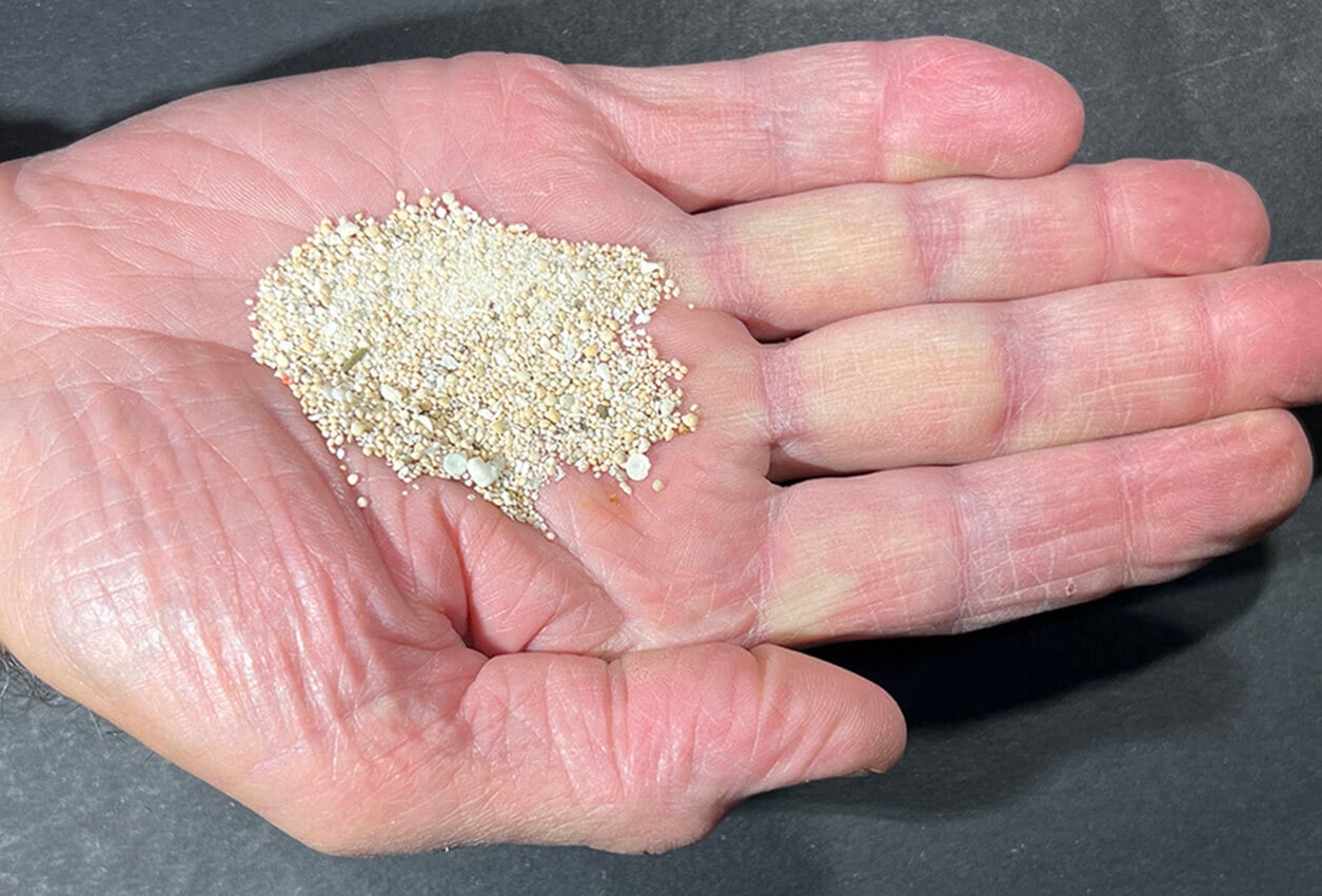
The powdery white sand at Nishi Beach is formed from eroded coral and microscopic marine organisms whose calcium carbonate exoskeletons are washed ashore.
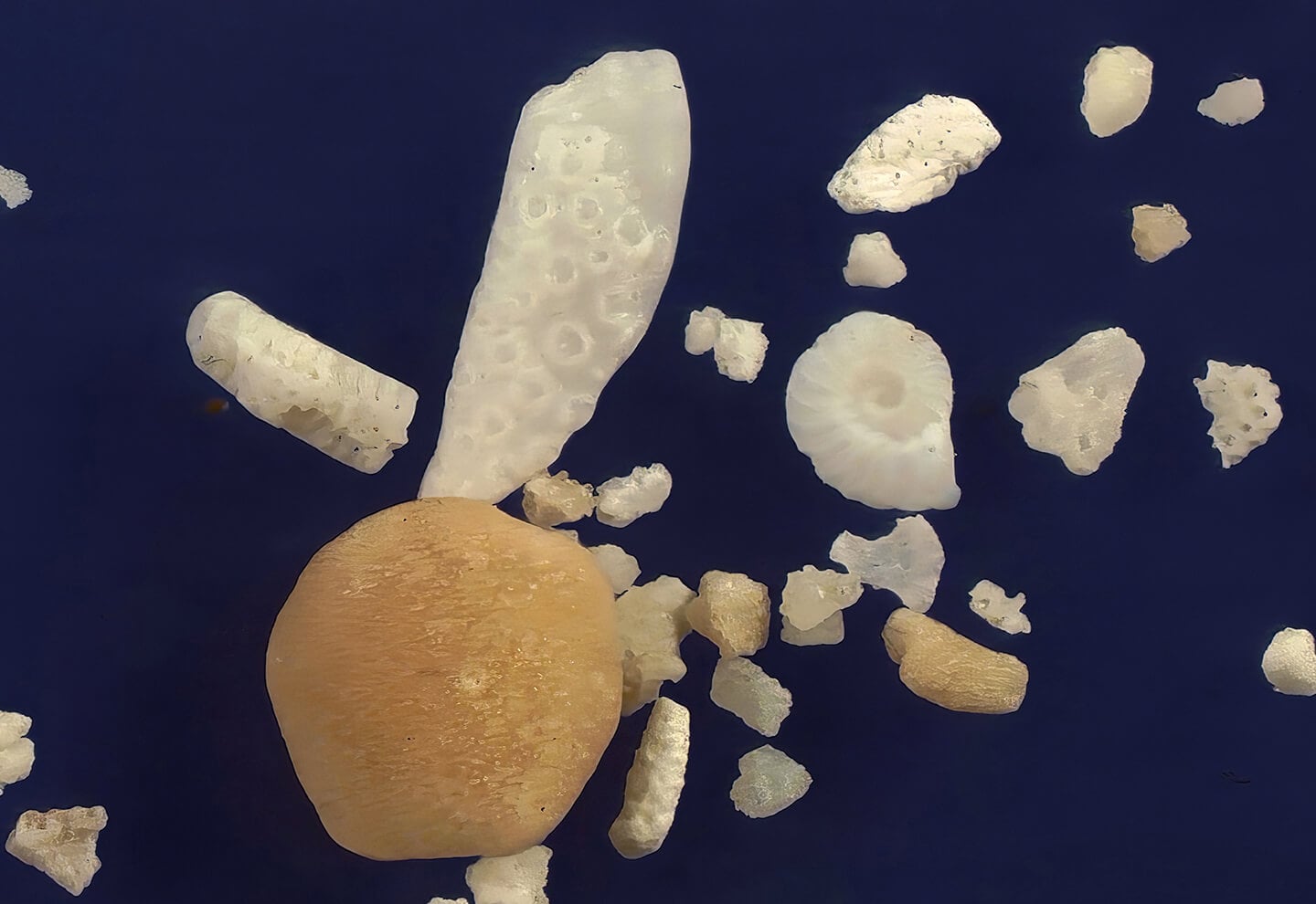
An unidentified elongated white grain with overlapping circular structure is above a round tan foram too worn to identify. The white coiled sand grain with subtle internal compartments just right of center is possibly an Operculina sp. foram.
Geographic Overview
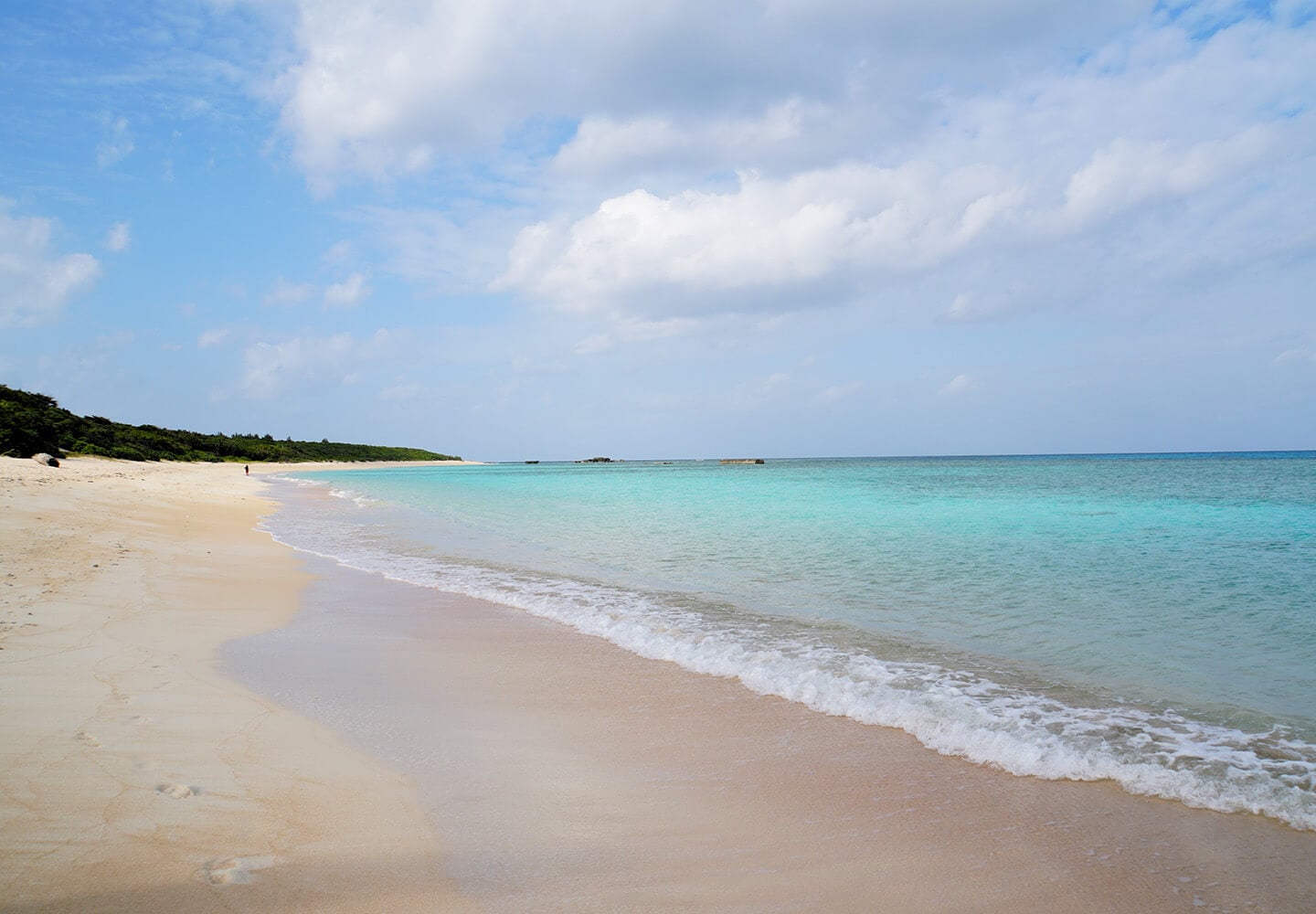
Nishi Beach (also known as Nishihama Beach) is a pristine, 1-kilometer stretch of white sand on Hateruma Island, Japan’s southernmost inhabited island. Renowned for its breathtaking turquoise waters, vibrant coral reefs, and diverse marine life, this beach captivates visitors with its stunning Hateruma Blue hue.
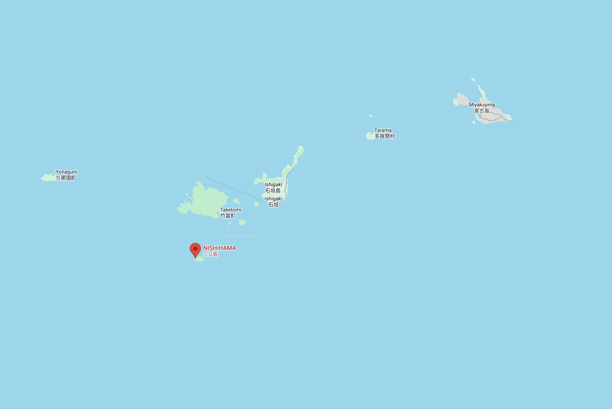
Sand Location
See where this sand sample was collected on Google Maps.
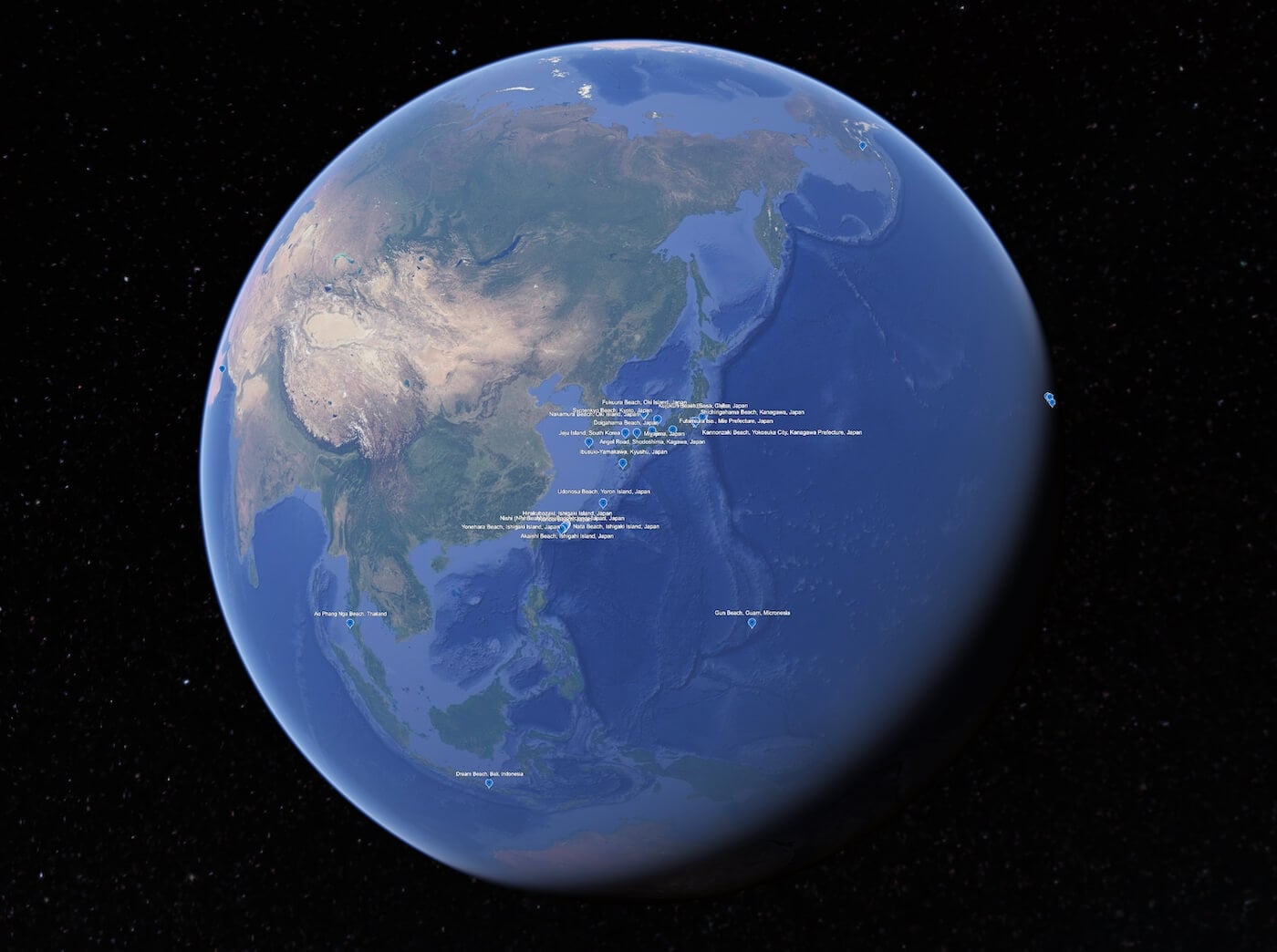
Virtual Sand Tour
Explore an interactive map of Nishi Beach on Google Earth.
Sand Gallery
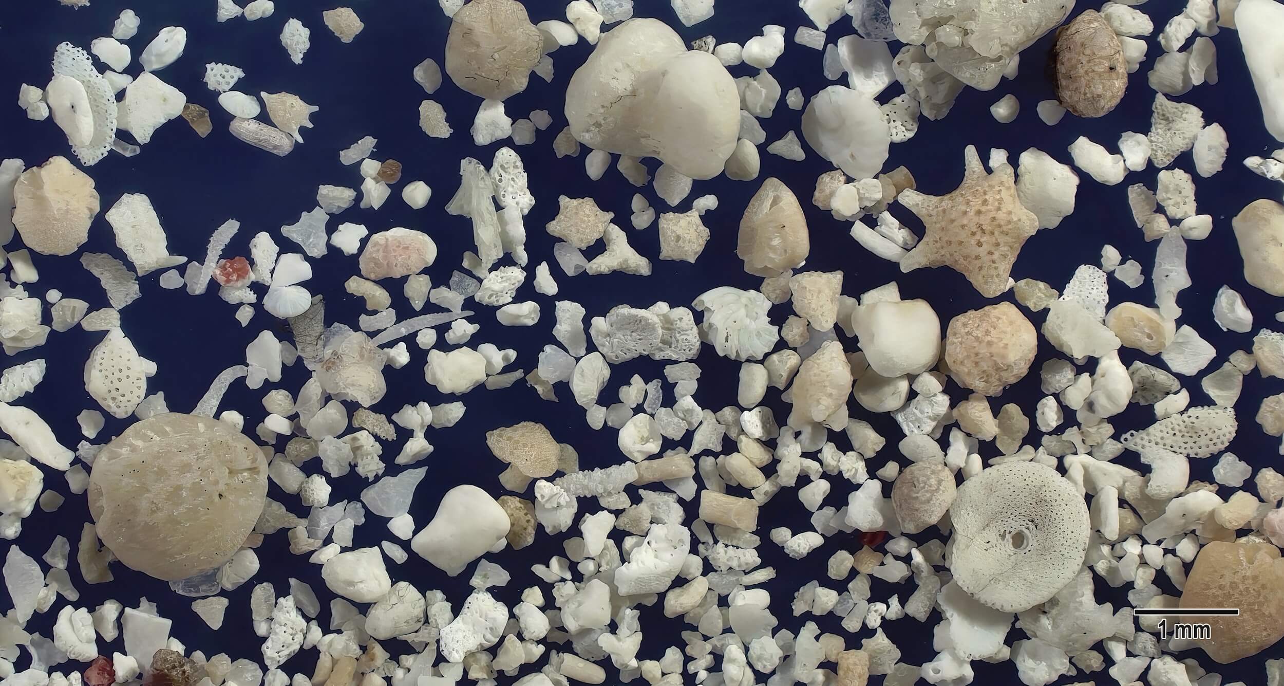
This low magnification mixture of large and small white and light tan sand grains includes several different forams. Larger forams include a round light tan foram with subtle parallel linear internal structure at the lower left, a relatively intact large round white foram with small parallel linear openings at the lower right (most likely Marginospora sp.), and brown spotted light tan grains, one with linear pointed extensions at the middle right (likely Baculogypsina sp.). A small white multichambered fan-shaped foram (possibly Polystomella sp.) is in the center and a subtly semitranslucent linear sclerite fragment surrounded by small marine sand fragments is left of center. The smooth surface large white sand grain at the top center is a fragment of coralline algae.
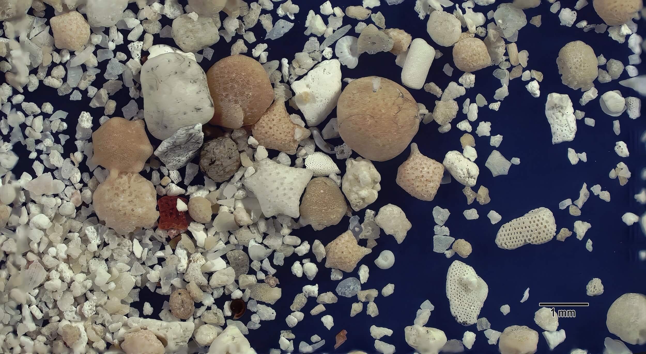
Against a dense background of marine sand grains too small to identify, a star-shaped white foram is just left of center and surrounded by large tan rounded sand grains represent different foraminifera species. White fragments with curved linear openings (likely Marginospora sp.) are near the bottom right. A short white rod fragment of coralline algae is at upper center just above a large tan foram.
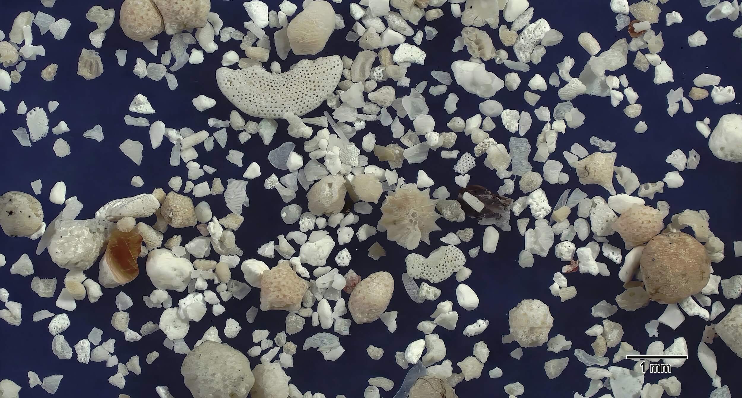
Most of the sand grains in this view are small white and semitranslucent fragments too small to identify. Larger sand grains represent a spectrum of foraminifera organisms including a sand grain with evenly spaced pointed peripheral exterior wall projections (possibly Neorotalia sp.) in the center, a fan-shaped fragment with parallel linear holes (may be Baculospina sp.) at upper left center, and a distribution of various other forams throughout this view. An unidentified brown fragment lies near the left middle edge and a black striped fragment is at right center.
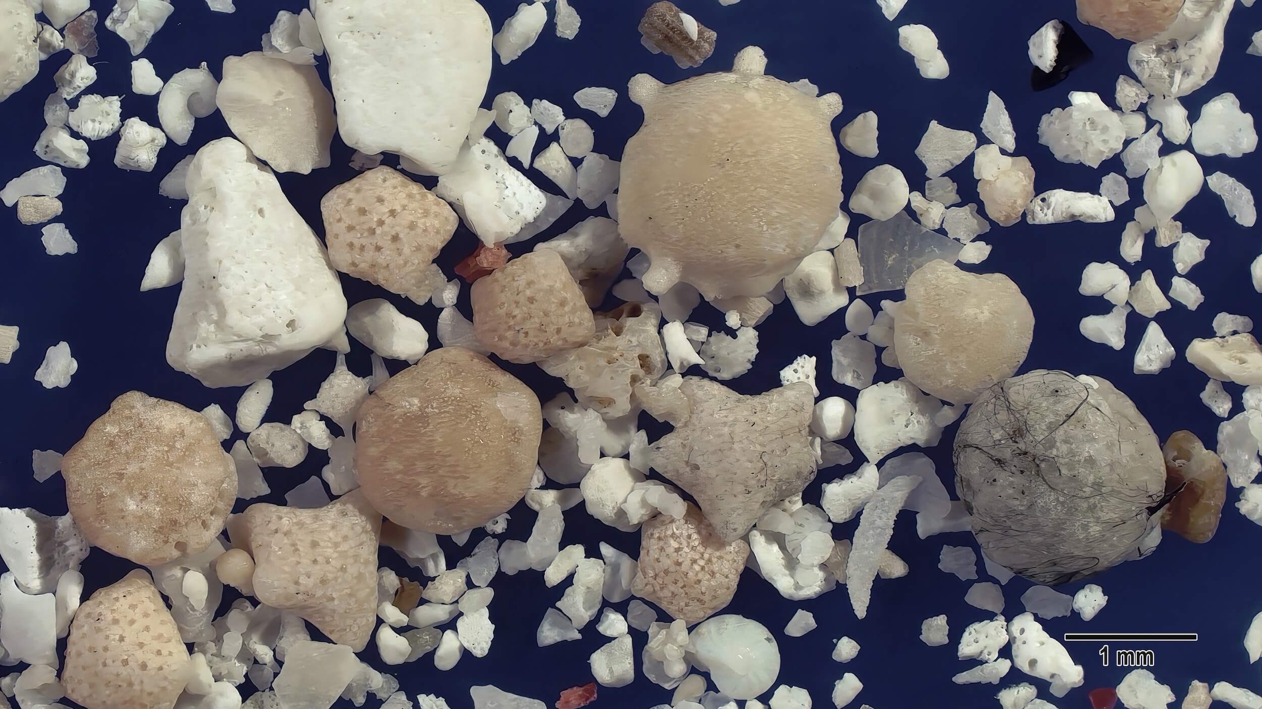
Against a background of white and off-white bits and pieces of marine shards, several large sand grains stand out, including two large white coralline algae fragments at the upper left, multiple tan and light tan smoothly worn grains representing multiple foraminifera species, and a light gray foram partly covered with black marine treads. A brown fragment of sea urchin spine is at the top center edge, a round white foram with barely visible compartments (possibly Operculina sp.) is at the bottom middle edge, and an elongated pale white sclerite spicule is near the bottom just right of center.
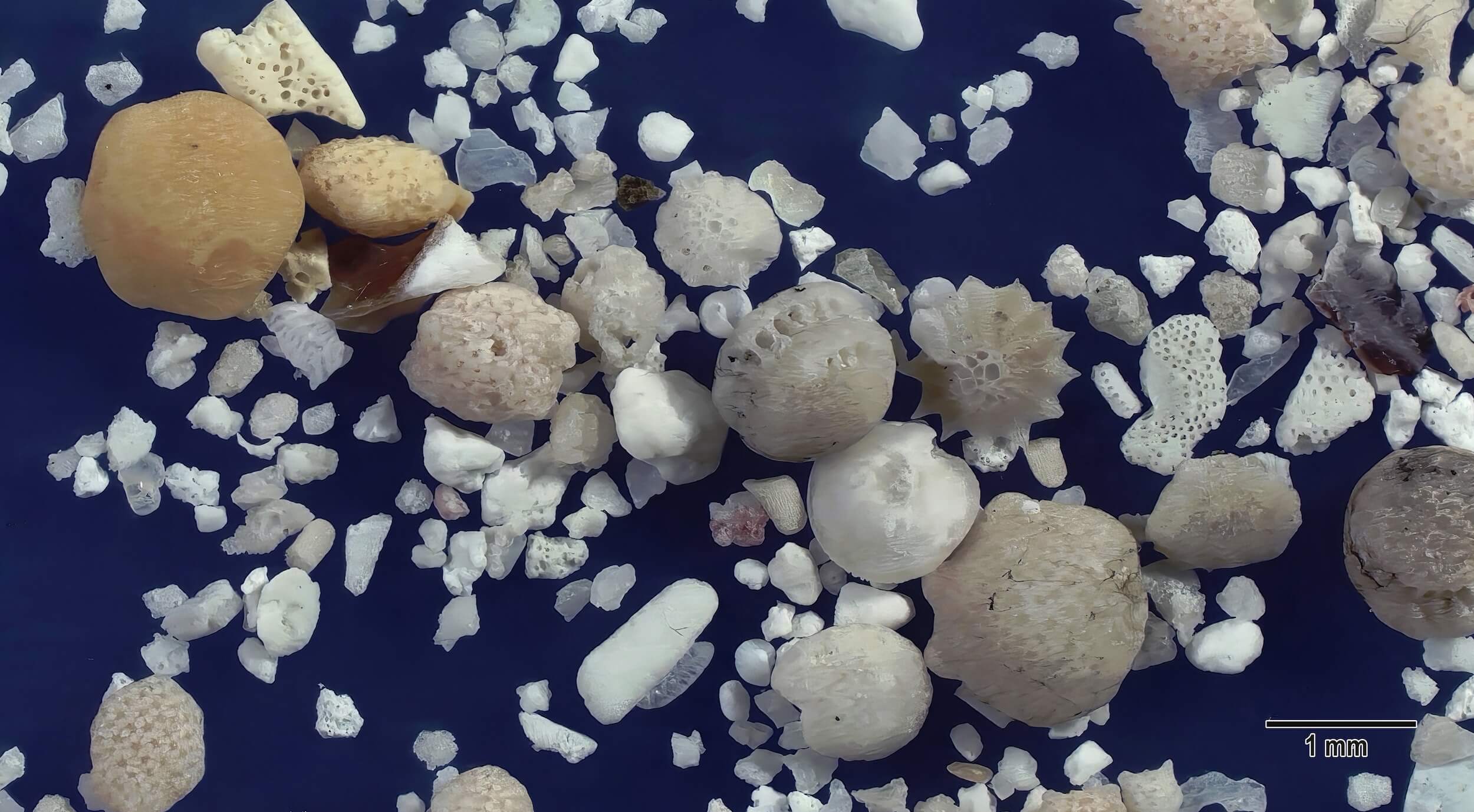
Several large tan and off-white foraminifera sand grains representing different species are present in this view along with a centrally located multipointed foram (likely Neorotalia sp). Two white fragments with tightly spaced linear openings just to the right of center are likely fragments of Marginospora sp.